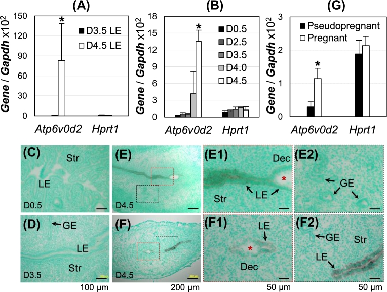
Figure 1.
Spatiotemporal expression of Atp6v0d2 in early pregnant WT mouse uterus. (A, B, G) Realtime PCR, normalized with Gapdh and Hprt1 as a second control; *P < 0.05; error bar: standard deviation; (C–F) in situ hybridization, counterstained with methyl green; D4.0, gestation day 4.0, about time for embryo attachment in mice. (A) Atp6v0d2 mRNA expression in D3.5 and D4.5 LE, N = 5–6. (B) Atp6v0d2 mRNA expression level in peri-implantation uterus, N = 4–6. (C–F) Localization of Atp6v0d2 mRNA in D0.5 (C), D3.5 (D), and D4.5 (E, F) uteri. E1 and E2, enlarged images from the red and black rectangle areas in E, respectively; F1 and F2, enlarged images from the red and black rectangle areas in F, respectively; red *, embryo; LE, uterine luminal epithelium; GE, uterine glandular epithelium; Str, stroma; Dec, decidual zone; scale bar, 100 μm (C, D), 200 μm (E, F), or 50 μm (E1, E2, F1, F2). (G) Atp6v0d2 mRNA expression in pseudopregnant and pregnant uterus at D3 22:00 h. N = 4–5.

