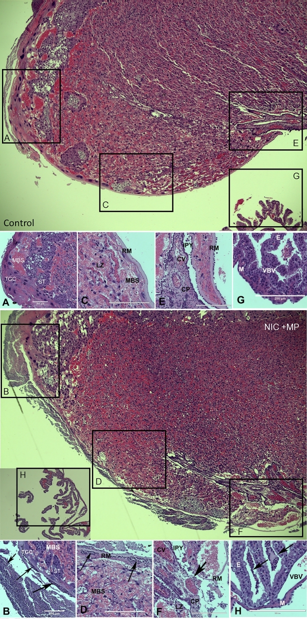
Figure 5.
Normal and representative severe histological lesions in the placenta. Low magnification images of control (top) and Nic + MP (bottom) placental disks with boxed areas indicating select sites subject to scoring: choriodecidua (boxes A and B), subchorion (boxes C and D), chorionic plate (boxes E and F), and visceral yolk sac (boxes G and H). Boxes correspond to the location of the structures shown in the high magnification image panels (control: A, C, E, G; Nic + MP: B, D, F, H). Arrows indicate neutrophils. Panel A shows normal choriodecidua. In Panel B, the decidua is obliterated by neutrophil infiltrates. Panel C shows normal subchorion. In Panel D, neutrophils infiltrate the maternal blood spaces at the periphery of the labyrinth zone. Panel E shows normal chorionic plate. Panel F shows epithelial effacement, neutrophil infiltration, and structural breakdown of the chorionic plate. Panel G shows normal visceral yolk sac. In Panel H, purulent exudate is seen among the endodermal villi of the visceral yolk sac. All images were obtained with an EVOS Auto FL imaging system (Life Technologies). Panels represent ×10 (A and B) or ×20 magnification. All scale bars are equivalent to 200 μm. Structural abbreviations: decidua (D), maternal blood sinus (MBS), trophoblastic giant cell (TGC), spongiotrophoblast (ST), labyrinth zone (LZ), Reichert's membrane (RM), intraplacental yolk sac (IPY), chorionic plate (CP), chorionic vessel (CV), endoderm (E), vitelline blood vessel (VBV), and mesothelium (M).

