Fig. 5.

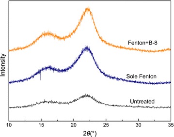
XRD patterns of the untreated and pretreated RS samples: the main diffraction peaks of 16.1° and 22.1° were assigned to the crystalline structures of cellulose I (101) and (002), respectively

XRD patterns of the untreated and pretreated RS samples: the main diffraction peaks of 16.1° and 22.1° were assigned to the crystalline structures of cellulose I (101) and (002), respectively