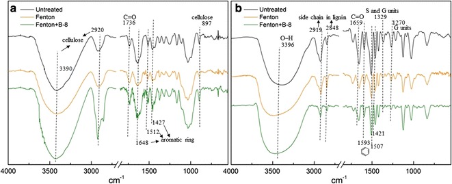
Fig. 6.

FTIR spectra of untreated and pretreated RS and lignin samples. a FTIR spectra of RS samples. The broad bands in the 3400–3300 cm−1 region, the band at 2920 and 897 cm−1 were cellulose characteristic peaks. The shoulder peak at 1736 cm−1 which associated with the carbonyl band (C=O) in lignin and/or hemicelluloses. The peaks at 1648, 1512 and 1427 cm−1 representing the lignin aromatic ring structure. b FTIR spectra of isolated lignin from RS samples. The shoulder peak at 3396 cm−1 derived from the O–H stretching. The absorption peaks at 2919 and 2848 cm−1 were the structure of the side chain in lignin. The peak at 1659 cm−1 derived from conjugated carbonyl (C=O). The characteristic peaks of the benzene ring skeleton appeared at 1593, 1507 and 1421 cm−1. The absorption peaks of S units and condensed G units at 1329 cm−1. G units C=O stretch at 1270 cm−1
