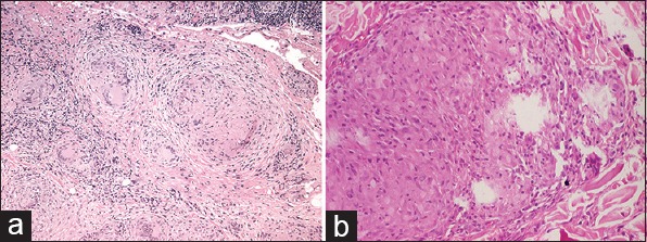
Figure 3.

(a) Multiple well-defined granulomas seen in the dermis having epithelioid histiocytes, multinucleated giant cells, and few lymphocytes (H and E, 10×). (b) Closer view showing epithelioid cell granuloma with histiocytes, epithelioid cells, and scattered lymphocytes (H and E, 40×)
