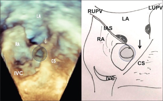
Figure 2.

Volume rendered three-dimensional echo after cropping the anterior half of both atria shows the enlarged coronary sinus ostium, through which the terminal unroofing is seen (circle). The pulmonary veins drain in the posterior wall of LA. The atrial septum is intact. RA: Right atrium, LA: Left atrium, CS: Coronary sinus, IVC: Inferior vena cava, LUPV: Left upper pulmonary vein, RUPV: Right upper pulmonary vein, IAS: Interatrial septum
