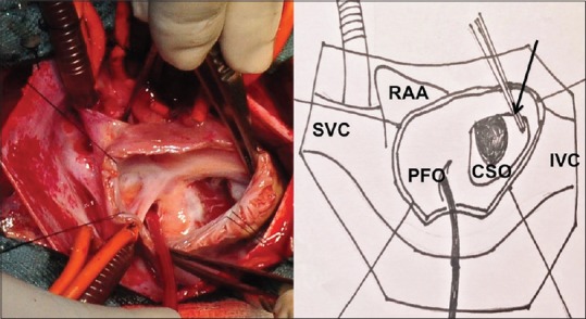
Figure 3.

Surgical view after opening the right atrium below the right atrial appendage and cannulation of the SVC and IVC, demonstrates terminal unroofing of the coronary sinus shown by arrow after inferiorly stretching its CSO with a forceps. A sucker is passed through the PFO into the left atrium. CSO: Coronary sinus ostium, PFO: Patent foramen ovale, IVC: Inferior vena cava, SVC: Superior vena cava, RAA: Right atrial appendage
