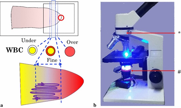
Fig. 1.

a Schema for the revised AO staining and slide examination. The AO droplet (indicated by the red open circle) on a cover slip is placed at the tip of the thin film. A typical scanning route to find parasites is indicated by the meandering line. b Picture of a blue LED lamp placed under the stage of a microscope. The LED lamp has a built-in shortpass filter (#) under its condenser lens and a longpass filter (*) is inserted in the microscope body
