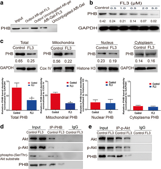
Fig. 2.

FL3 binds to PHB protein and inhibits the Akt/PHB interaction. a FL3-conjugated or unconjugated beads were incubated with total cell lysates from T24 cells. The eluted proteins were resolved on 10% SDS-PAGE gels for Western blotting with primary PHB antibody. b After incubation with indicated concentrations of FL3 for 24 h, total proteins of T24 cells were extracted and subjected to Western blot with primary PHB antibody. The Western blots have been quantified by densitometry and the quantitative values have been incorporated below the Western blot bands. c Subcellular fractions including cytoplasm, nucleus and mitochondria were isolated from the FL3-treated or control T24 cells. Then these fractions were lysed to obtain their total proteins, followed by Western blot analysis. Histograms show PHB protein intensity normalized to GAPDH, COX IV and Histone H3, respectively. The values represent the mean ± SD of three independent experiments. *P denotes < 0.05. The Western blots have been quantified by densitometry and the quantitative values have been incorporated below the Western blot bands. d After treatment with 0.5 μM FL3, total cell lysates of T24 cells were immunoprecipitated with primary PHB antibody. Then the eluted proteins were subjected to Western blot analysis with primary antibodies as indicated (left panel). e After treatment with 0.5 μM FL3, total cell lysates of T24 cells were immunoprecipitated with primary phospho-Akt (p-Akt) antibody. Then the eluted proteins were subjected to Western blot analysis with primary antibodies as indicated (left panel)
