
Fig. 3.
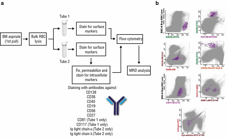
Minimal residual disease. A diagram of the protocol used to assess minimal residual disease shown in a. Bone marrow aspirates subjected to bulk red blood cell lysis and separated into 2 tubes. Each tube was incubated with an 8-marker antibody combination. Tube 1 was stained for surface markers only and tube 2 surface and intracellular markers. Minimal residual disease was then measured by flow cytometry merging data from each tube into one analysis. As shown in b plasma cells (PC) displayed a CD38high CD138int phenotype with low CD19 and CD45 expression indicative of an immature plasmablast (PC) population. By applying a cutoff value of 1 abnormal/clonal plasma cell per million nucleated event for MRD positivity (i.e., 10−6 sensitivity threshold of MRD positivity), the bone marrow aspirate tested MRD negative. BM bone marrow, RBCs red blood cells, MRD minimal residual disease