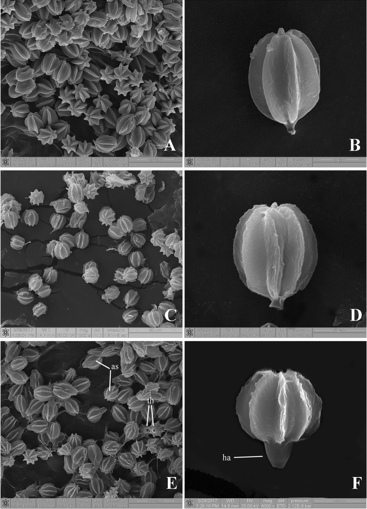
Figure 4.
Scanning electron micrographs of basidiospores A–B Rhodactina himalayensis (CMU25117) showing the basidiospores with 6–7 longitudinal ridges C–D Rhodactina incarnata (CMU25116, holotype) showing the basidiospores with 8–9 longitudinal ridges E–F Rhodactina rostratispora (O. Raspé 1055) showing the basidiospores with 8–9 longitudinal ridges, the wide and prominent hilar appendage (ha), a terminal hilum (th) and anastomosing ridges in some spores (as).

