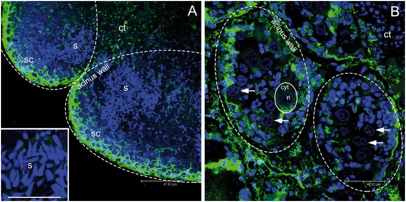
Figure 2.
Gonadic structure of Ruditapes philippinarum. (A) Two adjacent male acini at confocal microscope showing many spermatozoa (s; around 500 gametes per z-section). Magnification of sperm nuclei, in light blue, in the inset. (B) Two adjacent female acini (dashed ovals) at confocal microscope showing up to 10 eggs per section (1 egg is lined with a solid oval). The nucleus of some eggs is indicated by an arrow. (A, B scale bar = 47.62 µm; A inset scale bar = 16.26 µm). s = spermatozoa; sc = spermatogenic cells along the acinus wall; n = egg nucleus; cyt = egg cytoplasm; ct = connective tissue. Microtubules in green; TO-PRO3 nuclear dye in blue.

