Figure 3.
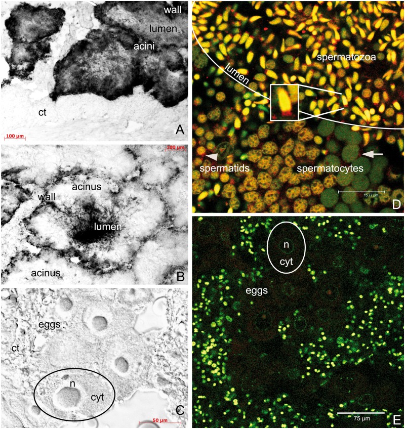
Localization of rphm21 products. (A–C) rphm21 transcript localization with in situ hybridization in male (A, B) and female (C) gonadic tissue of the Manila clam Ruditapes philippinarum. (A) Male immature acinus in which rphm21 riboprobe labels spermatogenic cells along the acinus wall (positive signal in black). (B) Male mature acinus in which spermatozoa stored in the acinus lumen are deeply stained with rphm21 riboprobe. (A, B scale bars = 100 µm). (C) No staining is present in eggs (C scale bar = 50 µm). n = egg nucleus; cyt = egg cytoplasm; ct = connective tissue. (D, E) RPHM21 protein localization in male and female gonadic tissue, respectively. (D) In the male gonad, anti-RPHM21 (in red) labeled a clear spot at 1 side of the nucleus of spermatocytes and spermatids (arrow and arrowhead, respectively). Anti-RPHM21 staining is strong in mature spermatozoa in the acinus lumen in both mitochondrial midpiece and nucleus (yellow due to the colocalization of the nuclear dye, in green, and the antibody, in red). Initially, in spermatocytes the nuclei are visible in green, and no RPHM21 appears to be stored in the nucleus; in spermatids the nuclei are brownish as indicating RPHM21 storing (clearly seen in the bottom of the image going from the right to the left). (D scale bar = 15.77 µm). (E) Eggs do not show anti-RPHM21 detectable staining. The smaller nuclei of somatic cells surrounding acini are also visible. (E scale bars = 75 µm). RPHM21 immunolabeling in red; TO-PRO3 nuclear dye in green.
A-C: optical microscope. D, E: confocal microscope.

