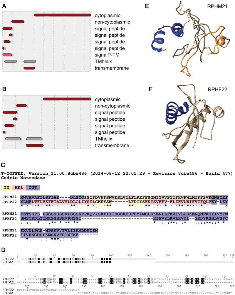
Figure 4.
Structural analysis of RPHM21 and RPHF22. (A, B) Protein domains detected with InterProScan 5: RPHM21 (A) and RPHF22 (B) both show transmembrane domains in their N-terminus, whereas the C-terminus is cytoplasmic. (C) Similarities in domain localization detected with TM-COFFEE alignment. (D) HMMER detected good alignment of profile HMMs in correspondence of the transmembrane domains. (E, F) 3D models of RPHM21 (E) and RPHF22 (F) obtained using structures predicted by I-TASSER.

