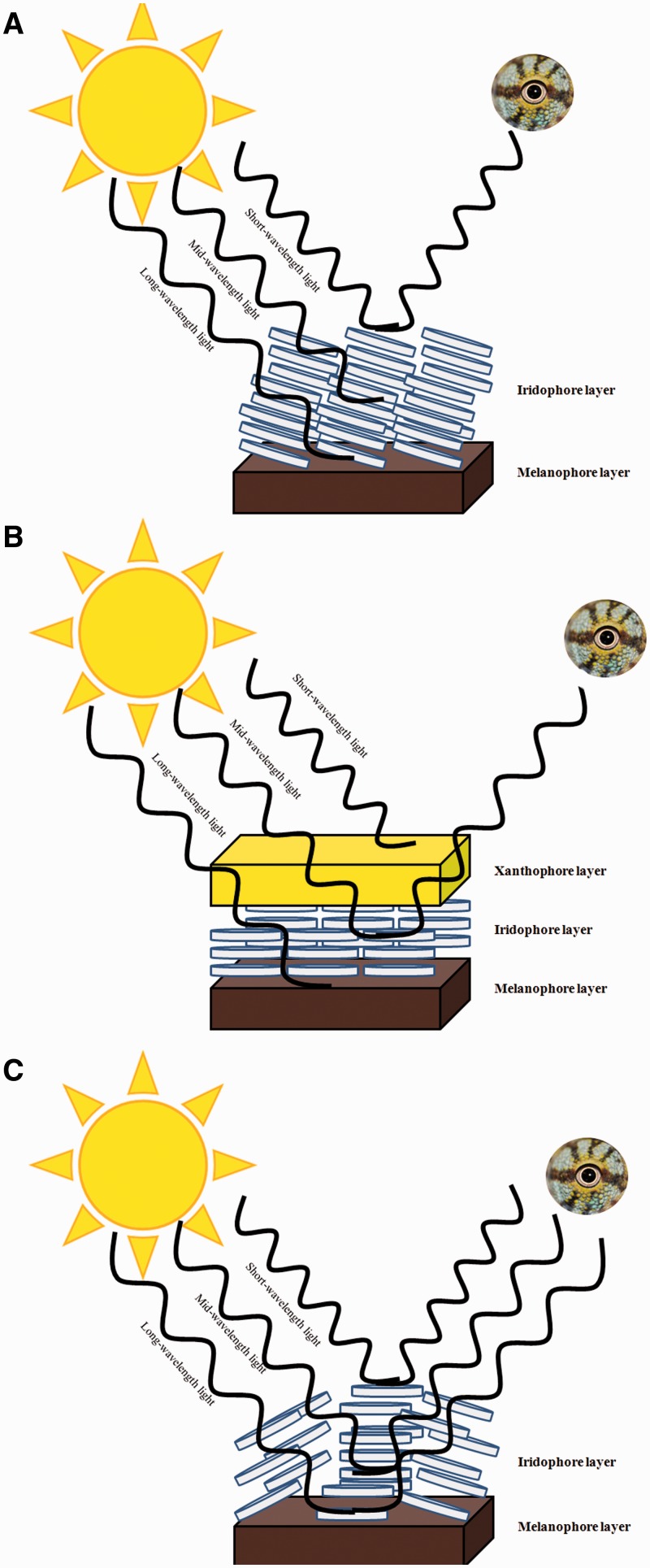
Figure 2.
Schematic representation of the potential contributions of the different elements of the dermal chromatophore unit to reflected light (i.e., color and brightness). Light from an external source (e.g., the sun) interacts with the dermal chromatophore, which influences the wavelengths of light transmitted from the body (and which become visible to external viewers).
Notes: (A) Blue color can be attained when reflecting platelets in the iridophore layer selectively reflect short wavelengths of light and longer wavelengths of light are absorbed by the melanin-containing melanophore layer. (B) Green or yellowish-green is reflected when the xanthophore layer absorbs short-wavelengths of light, the platelets in the iridophore layer reflect medium wavelengths of light, and the melanophore absorbs longer wavelengths of light. (C) White light is reflected when iridophore platelets reflect or scatter all incoming wavelengths of light. Figure after Bagnara and Hadley (1973).

