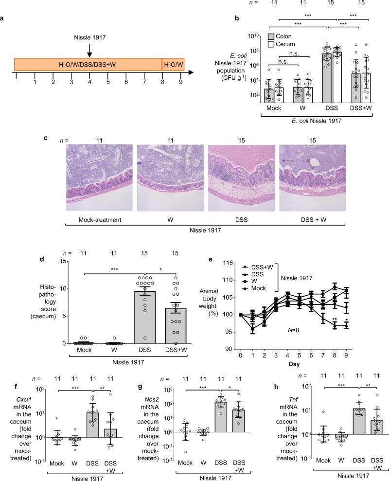
Extended Data Figure 3. Impact of tungstate treatment on mice experimentally colonized with E. coli Nissle 1917.
Groups of conventionally-raised C57BL/6 mice were orally inoculated with the E. coli Nissle 1917 wild-type strain and treated with 0.2 % sodium tungstate (W), dextran sulfate sodium (DSS), DSS and sodium tungstate, or left untreated (mock) for 9 days. a, Schematic representation of colitis model used in this figure. b, Bacterial load in the cecum (white bars) and colon content (black bars). c–d Formalin-fixed, hematoxylin and eosin-stained sections of the cecum were scored for the presence of inflammatory lesions. c, Representative images of stained cecal sections. d, Cumulative histopathology score for the cecum tissue; bars represent means ± standard error and each dot represents one animal. b–d, Mock and W (n=11 per group), DSS and DSS+W (n=15 per group). e, Animal body weight, n=8 per group. f–h The transcription of the inflammatory marker genes Cxcl1 (f), Nos2, (g) and Tnf (h) in the cecal mucosa was determined by RT-qPCR, n=11 per group.
Unless noted otherwise, bars represent the geometric mean ± geometric standard deviation. *, P < 0.05; **, P < 0.01; ***, P < 0.001. n.s, not significant.

