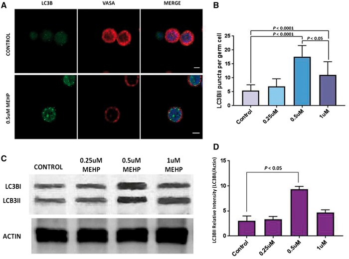
Figure 2:
autophagy in porcine germ cells exposed to increasing concentrations of MEHP. (A) Double immunolabeling of LC3BII and VASA in porcine germ cells. LC3BII puncta were increased in MEHP exposed cells (inferior panels) compared to the control cells (control panels). Nuclei are stained with DAPI. Scale= 5 μm. (B) Number of LC3BII puncta per cell is shown, for four different MEHP concentrations. 60 cells per treatment were analyzed, and 3 independent experiments were performed. The data is mean ± SD, comparison made by one-way ANOVA. (C) Immunoblot of lysates from the porcine germ cells exposed to increasing concentrations of MEHP. (D) The relative intensity of LC3BII bands were calculated from the scanned intensities of bands of 3 independent experiments exemplified by the Western blot shown in (C). Data is mean ± SD, comparisons made by one-way ANOVA.

