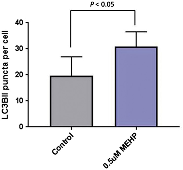
Figure 3:

autophagic flux in porcine germ cells exposed to MEHP. Cells exposed to 0.5 μM MEHP were treated with bafilomycin A1 for 4 h previous to collection. LC3BII puncta per cell are shown. Sixty cells per treatment were analyzed from 3 independent experiments. Data shown is mean ± SEM, analyzed by unpaired t-test.
