Figure 6:
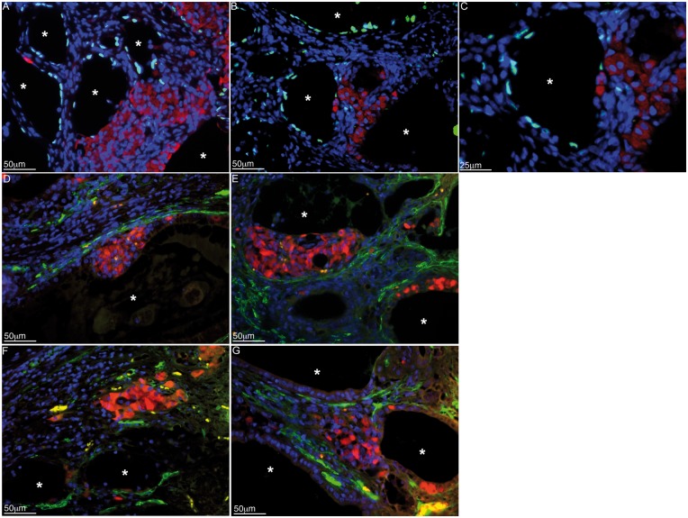
Immunolocalization of islets, SCs and myoid cells in the grafts. Primary mouse SCs were isolated from BALB/c mice (8–10 days old; Charles Rivers Laboratories; Wilmigton, MA) and cultured in vitro as described in Fig. 1. Islets were isolated from male BALB/c mice (6–8 weeks old; Charles Rivers Laboratories) by collagenase digestion of the pancreas and Ficoll density gradient purification. The isolated islets were cultured in nontreated petri dishes in Ham’s F10 media with supplements at 37 °C. Three million BALB/c SCs were co-transplanted with 500 BALB/c islets underneath the kidney capsule of diabetic C3H mice as allografts. The graft bearing kidneys were collected from normoglycemic mice at day 102 post-transplantation. The tissue sections were double immunostained for WT1 (SC marker, green, A–C) or smooth muscle alpha-actin (myoid cell marker, green, D–G) and insulin (islet cell marker, red, A–G; 1:1000 dilution; Dako/Agilent Technologies). Sections were counterstained with DAPI to detect cell nuclei (blue color, A–G; ThermoFisher Scientific). Asterisks represent SC tubules.

