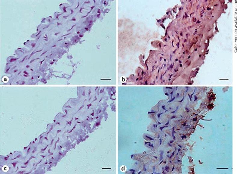
Fig. 5.
Immunostaining for IP3 receptor expression in C57BL/6 (a, b) and ApoE−/− (c, d) aortic rings. Aortae were removed from ApoE−/− mice on the diet for 4 months and age-matched C57 mice. No staining was visible in arteries in which the primary antibody was omitted (a, c). IP3 receptor was diffusely expressed throughout the medial layer in C57BL/6 mice (b), with a clear reduction in receptor expression in the high-fat-fed ApoE−/− mice (d). Representative images are shown, with a minimum of 4 arteries studied for each group. Scale bars, 10 μm.

