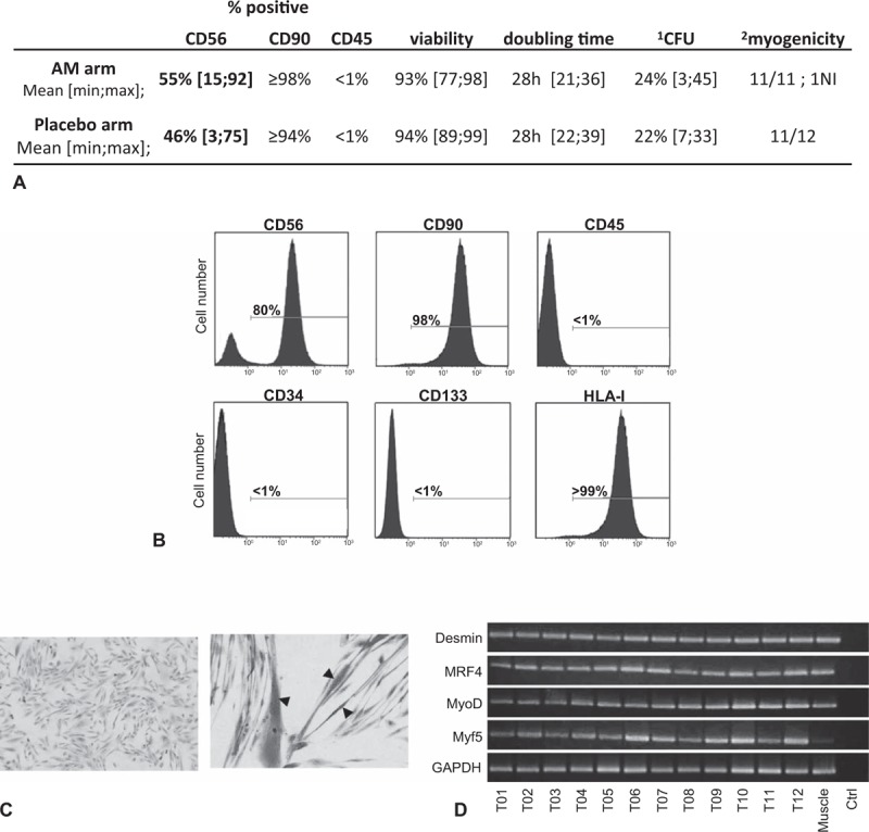
FIGURE 2.

Characterization of myoblast preparation. A, Immunophenotype and growth characteristics of myoblast preparations. B, Example of immunophenotype for myoblast preparation (T02): flow cytometry analysis after staining with anti-CD56, anti-CD90, anti-CD45, anti-CD34, anti-CD133 and anti-HLA-I monoclonal antibodies. Complete phenotypic results appear in supplemental Table 1. C, Example of undifferentiated myoblasts for P07 (left, magnification 100×) and myogenic differentiation (right, magnification 200×) in appropriate culture medium. Arrowhead, multinucleated fiber. Giemsa staining. D, RT-PCR analysis of desmin and myogenic factor (MRF4, MyoD, and Myf5) gene expression. cDNAs were from the 12 different myoblast preparations from the treated arm and from 1 normal muscle sample. Control is RT-PCR without RNA template at the RT step. ∗CFU, colony-forming units (20 or 40 cells/well, 6 wells); †Capacity of clones to give rise to plurinucleated myotubes. AM indicates autologous myoblasts; NI, not interpretable.
