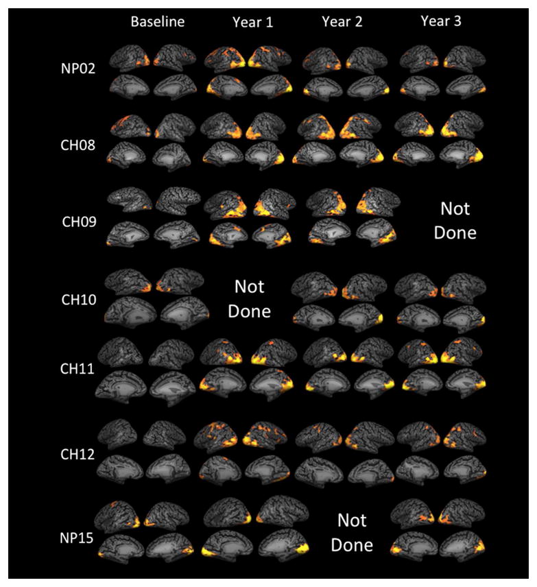
Figure 1. Longitudinal fMRI results for individual RPE65 participants superimposed on their inflated cortex.

fMRI results for 7 RPE65 subjects at baseline, one year, two years, and three years post retinal gene augmentation therapy are presented in Figure 1. Cortical activations are assessed in response to the stimulation of the newly treated eye for both the high and medium contrast stimuli.19 Results are presented for the left eye for all but one participant (CH09) who received re-administration to his right eye. fMRI results for all subjects were corrected for multiple comparisons using false discover rate (fdr < 5%) and a corrected P < 0.004, with a continuous connected area threshold (cca ≥ 100 mm2) that further controls for multiple comparison type 1 error.19, 23 In order to show cortical activations at baseline, fMRI results required considerably relaxed statistical threshold of an uncorrected P<0.05 and cca ≥ 50 except for NP15 and NP02. Using the above statistics, statistically significant areas of cortical activations are shown as yellow/orange clusters overlaid onto the medial and lateral representations of inflated cortex for each individual subjects. Overall comparison of the baseline and three year follow up fMRI results show noticeable increase in widespread bilateral activations in all areas of visual cortex extending from medial to lateral and posterior to anterior aspects of the occipital cortex one year after gene therapy for majority of the subjects. As shown in Figure 1, activation pattern vary for individual subject depending on his/he age and disease progression or other reasons. Changes in cortical activations over time for baseline and one year after retinal gene therapy using responses to only the high contrast stimuli (here we used responses to the high and medium contrasts) were reported in a recent report by Bennett et al. (2016).
