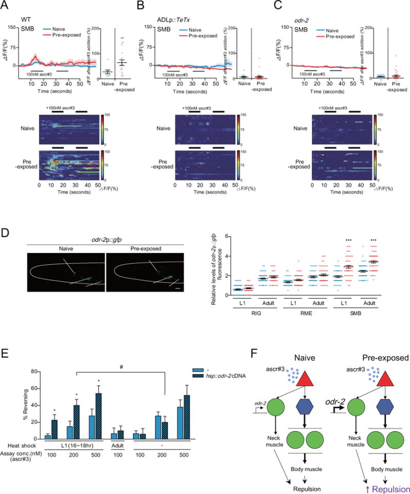
Figure 4. The SMB motor neurons mediate enhanced ascr#3 avoidance in ascr#3 imprinted animals.

A–C. Ca2+ transients of the SMB neurons in response to 100 nM ascr#3 in wild-type animals (A), wild-type animals expressing ADLp∷TeTx (B) or odr-2 mutants (C). The average traces of Ca2+ responses during two pulses of 100 nM ascr#3 (left), heat map of individual Ca2+ responses to ascr#3 (bottom), and the maximum value of Ca2+ responses to 100 nM ascr#3 (right) in naive (blue) or pre-exposed animals [10] are shown. ** indicates different from naive at p<0.01 by Student’s t-test. n=8–30 each.
D. Representative images (left) and GFP quantification (right) of odr-2p∷gfp expression in RIG, RME, and SMB of naive (shown in blue) and pre-exposed (shown in red) animals at the L1 or adult stages are shown. The images were taken of adults. *** indicates different from WT at p<0.001 by one-way ANOVA with Bonferroni’s post hoc test. n=50–60 each. Scale bar: 10 m.
E. Percentage of reversal of adult transgenic animals expressing hsp∷odr-2 cDNA under heat shock at the L1 and adult stages. * indicates different from hsp∷odr-2 cDNA (−) and # indicates different from no heat shock at p<0.05 by Student’s t-test. n=50–70 each. (A–E) Error bars represent SEM.
F. Model for circuit mechanisms underlying ascr#3 avoidance in naive or ascr#3 imprinted animals. In naive animals, ADL detects ascr#3 and transmits the signals to the post-synaptic AVA command interneurons and downstream VA/DA motor neurons. In pre-exposed animals, the increased expression of odr-2 in SMB initiates synaptic transmission from ADL to SMB that mediate ascr#3 signal transmission from ADL not only to AVA but SMB. SMB activation may serve to sensitize background for backward movement via excitation of head muscles.
See also Figure S4.
