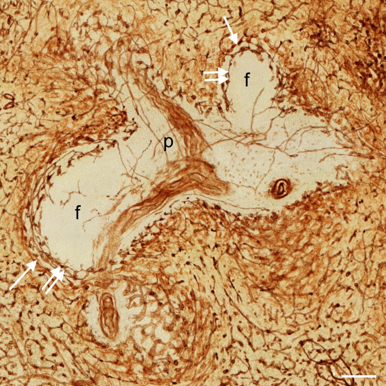
Fig 3. Visualisation of capillary networks centered on PALS and follicles.
Distribution of CD34 in an overlay of 24 registered sections of ROI 4 showing a PALS (P) with a branching central artery and two sectioned follicles (f). There is a clear difference in the localisation of the perifollicular capillary network (double arrows) and the perifollicular sinus network (arrows). Internal capillaries of the PALS appear to run in the adventitia of the central artery forming vasa vasorum. Scale bar = 100 μm.

