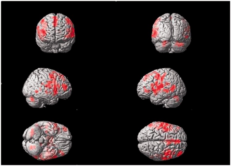
Figure 2.
Areas of brain activation induced by acupuncture stimulation of the right Tongli (HT5) acupoint. The results from functional magnetic resonance imaging were surface-rendered onto a canonical brain. The red areas represent all voxels that were significant at P < 0.005. The colour version of this figure is available at: http://imr.sagepub.com.

