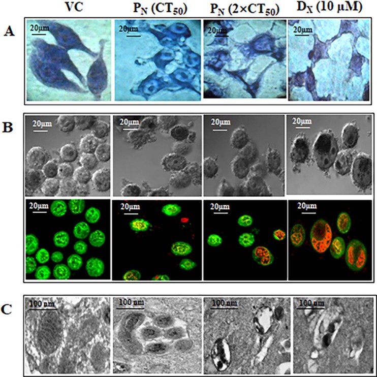
Fig 2.
Representative photomicrographs of morphology of HeLa cells stained with (A) Giemsa (B) Acridine orange with ethidium bromide, fluorescent microscopy showing intracellular entry of PN at 48h and visualized at 40 × magnifications. (C) Electron micrograph of ultra-thin section of VC, PN (CT50 and 2×CT50) and DX treated HeLa cells are shown at 48h and visualized at 15000× magnifications. Mitochondria show an interconnected network structure with numerous regularly arranged cristae, i.e., intact/condensed structures in VC compared to PN and DX treated cells. PN: Pinostrobin, DX: Doxorubicin, VC: Vehicle control.

