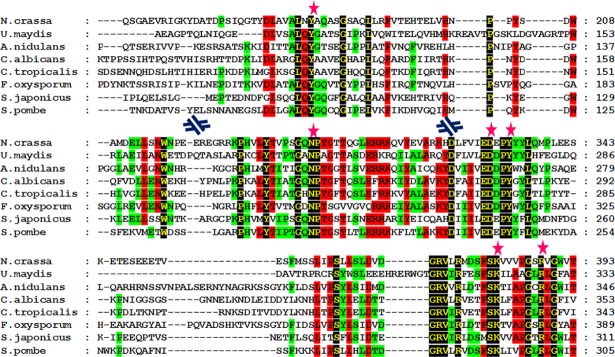
Fig 1. Multiple sequence alignment of tryptophan aminotransferase homologs from different fungal species.
Residues with 100% identity are shown in yellow font with a black background, residues with 75% identity are shown in black font with a red background, and residues with 50% identity are shown in black font with a green background. PLP binding residues are marked with a pink star at the top. Sequences are presented in a discontinuous fashion. Blue discontinuous symbols are shown when there is a discontinuity of the sequence.

