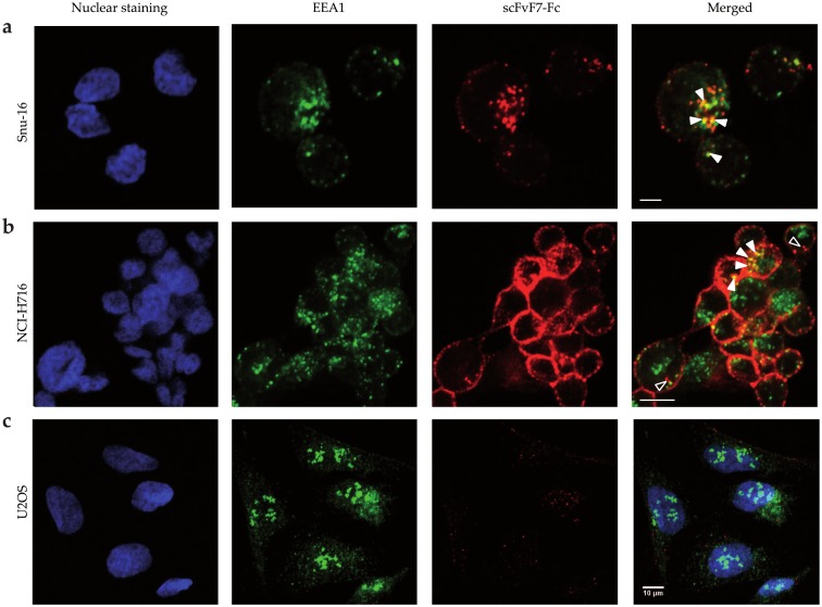
Fig 6. Localization of scFvF7-Fc in FGFR2-positive and FGFR2-negative cells.
Fluorescence was detected in fixed and permeabilized (a) Snu-16, (b) NCI-H716, (c) U2OS cells. scFvF7-Fc was stained red with DyLight550. EEA1 was labeled with anti-EEA1 antibody detected with Alexa Fluor 488–conjugated secondary antibody (green staining). Nuclei were stained with DAPI (blue). The scale bar in all panels represents 10 μm.

