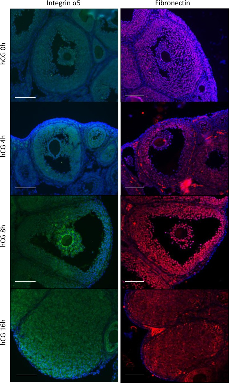
Fig 1. Expression of fibronectin and integrins in the mouse ovary during ovulation.
Localization of fibronectin and integrin α5 in the mouse ovary was detected by immunofluorescence staining (n = 3 mice in each time point). Proteins were visualized with Cy3 (fibronectin) and FITC (integrin α5). The nucleus was counterstained with DAPI. Scale bar is 100 μm.

