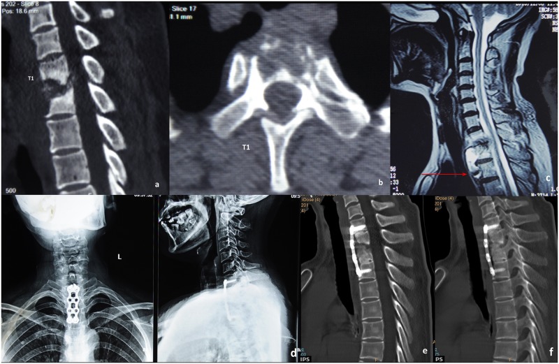
Fig 2. A typical case for group A.
A 56-year-old patient’s preoperative CT scanning shows destructive segments located at the T1 segments with corrasion of the T1 vertebra (a-b). Preoperative sagittal MRI shows that the tuberculosis focus is located higher than the suprasternal notch level (c). One-week postoperative X-ray image shows internal fixation in good position (d). Six-month postoperative CT scanning reveals no cervicothoracic anterior graft fusion yet (e). Three-year postoperative CT scanning reveals cervicothoracic anterior graft fusion (f).

