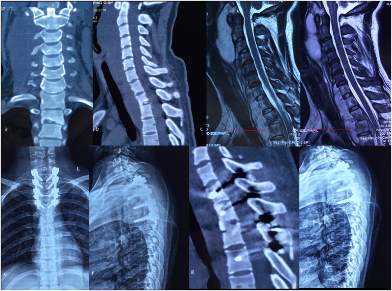
Fig 4. A typical case for group B and C.
A 27-year-old patient’s preoperative CT scanning shows destructive segments located at the T2/3 segments (a-b). Preoperative MRI shows a huge paravertebral abscess located in front of the vertebral bodies and the compression of the spinal cord, while the tuberculosis focus lies exactly on the suprasternal notch level (c-d). Two-week postoperative antero-posterior and lateral plain radiograph shows the internal instruments in a satisfactory position (e-f). Four-year postoperative CT scanning demonstrates that the cervicothoracic fusion is consolidated completely (g). Six-year postoperative lateral plain radiograph shows no instrumentation loosening, migration or breakage (h).

