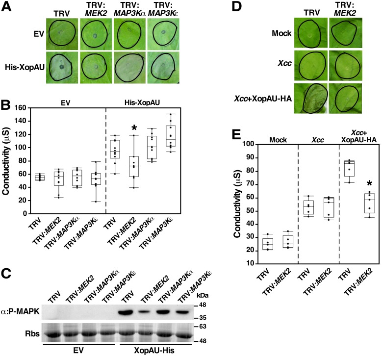
Fig 7. Silencing of MEK2 in N. benthamiana reduces XopAU-induced cell death.
N. benthamiana plants were infected with TRV, TRV:MEK2, TRV:MAP3Kα and TRV:MAP3Kε. In (A), (B), and (C), leaves of silenced plants were inoculated with Agrobacterium (OD600 = 0.02) carrying a vector either empty (EV) or for expression of His-XopAU from an estradiol-inducible system, and treated with 17β-estradiol 24 h later. (A) Photographs of inoculated areas at 36 h after 17β-estradiol application. (B) Electrolyte leakage at 24 h after 17β-estradiol application. (C) Total proteins were extracted at 12 h after 17β-estradiol application and samples were immunoblotted with α:P-MAPK antibodies. Rbs, Rubisco loading control stained by Ponceau S. In (D) and (E), leaves of silenced plants were inoculated with mock or suspensions (5 x 10⁷ CFU/ml) of Xcc carrying a vector either empty (EV) or for expression of XopAU-HA. (D) Photographs of inoculated areas at 48 h after Xcc inoculation. (E) Electrolyte leakage at 24 h after Xcc inoculation. In (B) and (E), box plots display 25th, 50th (middle line) and 75th percentiles. (in B, n = 10; in E, n = 5 or 7). Asterisks indicate a significant difference (Mann-Whitney U test, p value <0.05) relative to TRV empty control. Experiments were repeated three times with similar results.

