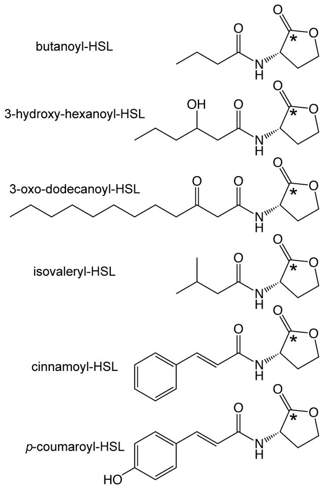
Figure 1.

Examples of AHL QS signal structures. The top three compounds are representative of the ‘typical’ AHL molecule, which has a side chain derived from fatty acid biosynthesis (acyl-acyl carrier protein substrates). The bottom three compounds are the more recently discovered AHL signals derived from branched chain amino acid biosynthesis (isovaleryl-HSL) and aromatic acid degradation (cinnamoyl-HSL, p-coumaroyl-HSL), which utilize acyl-coenzyme A (CoA)-linked substrates [6,9]. The asterisk indicates the location of the 14C-label incorporated in the 14C-AHL during the radiolabel assay protocol described in this chapter.
