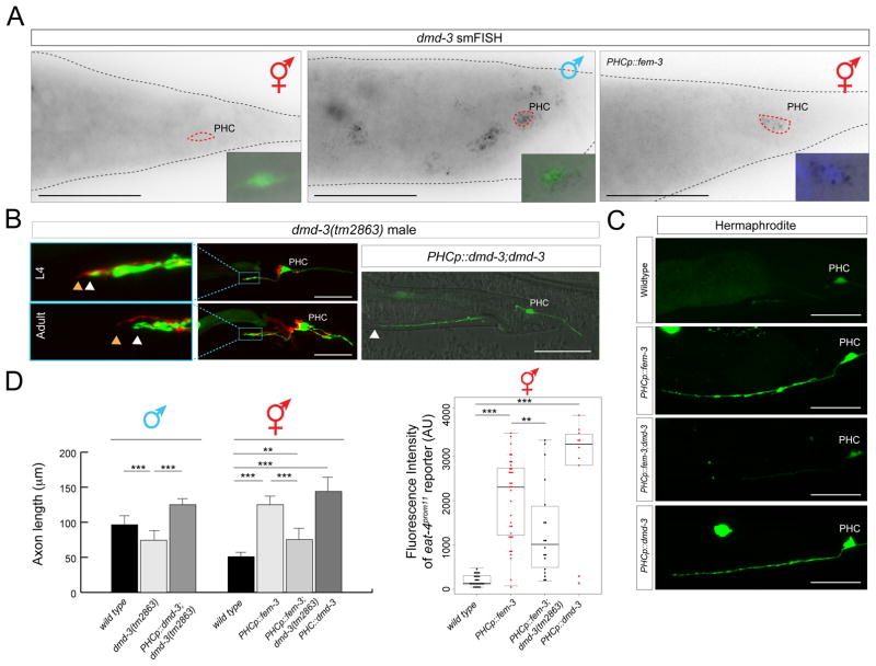
Fig. 6. dmd-3 expression and function.
A: smFISH analysis of endogenous dmd-3 expression in young adult animals. No expression is observed in hermaphrodites; in males, expression is observed in multiple cells including PHC, marked with the transgenic array otIs520. Masculinization of PHC (via PHCp::fem-3) in otherwise hermaphroditic animals activates dmd-3 transcription. The inset shows the PHC nucleus stained with DAPI. More than 10 animals were scored for presence of dots in each condition and all animals showed similar staining patterns relative to one another. See Fig. S2 for dmd-3 smFISH probe specificity. Scale bar: 50 μm.
B: PHC axons fail to extend in dmd-3(tm2863) mutant males and these defects are rescued by PHC-specific expression of dmd-3 [oExt6908 (eat-4p11Δ11::dmd-3)]. Scale bars: 50 μm. Wildtype and PHCp::fem-3 data showing in this plot is the same as in Fig. 2B for the adult stage. See panel D for quantification.
C: Ectopic expression of dmd-3 in PHC of hermaphrodites is sufficient to scale eat-4/VGLUT expression (4th panel) and dmd-3 is required in hermaphrodites for the axon extension conferred by masculinization of PHC (2nd and 3rd panel). Transgenic array names: otEx6879, otEx6880 (eat-4p11Δ11::fem-3); otEx6908(eat-4p11Δ11::dmd-3). See panel D for quantification.
D: Quantification of the axon extension and eat-4/VGLUT scaling in the different conditions showed in previous panels. Significance was calculated using student t-test, ***P < 0.005, **P < 0.05.

