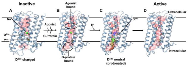
Figure 6. Proposed role of Na+ translocation in GPCR activation.
Key checkpoints during the transition from the inactive (A) to active (D) state of the receptor. (A) The initial, inactive receptor conformation shows no bound agonist or G-protein, and displays a Na+ ion bound in a pocket which is sealed towards the cytosol by a hydrophobic layer around Y7.53. (B) G-protein and agonist bind to the receptor (in undetermined order), leading to the formation of a continuous water channel across the GPCR. The increased mobility of the Na+ ion results in a pKa shift and subsequent protonation of D2.50. (C) Neutralization of D2.50 and the presence of the hydrated pathway facilitate transfer of Na+ to the intracellular side, driven by the transmembrane Na+ gradient and the negative cytoplasmic membrane voltage. (D) The expulsion of Na+ towards the cytosol results in a prolonged active state of the receptor.

