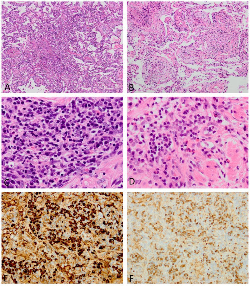
Figure 4. Histopathologic features of lungs involved in IgG4-RD.
(A) The inflammatory process infiltrates the alveolar interstitium. (B) An organizing pneumonia pattern is focally seen. (C) Although many plasma cells are observed, intervening lymphocytes are also present. (D) Occasional eosinophils are identified. (E and F) The absolute number of IgG4-positive plasma cells and ratio of IgG4/IgG-positive plasma cells are both increased (E, IgG4 immunostaining; F, IgG immunostaining). Images E and F were taken at almost the same fields.

