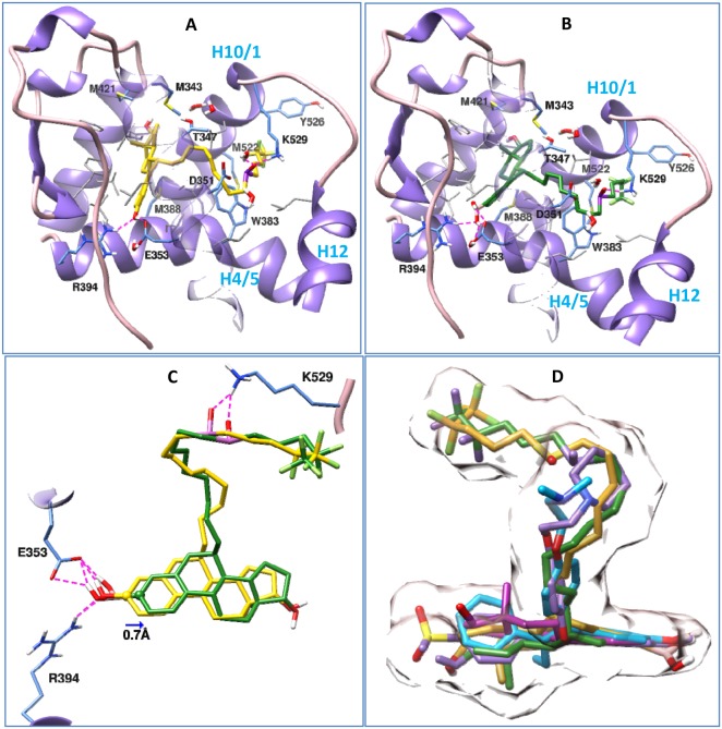
Figure 2. Binding postures of Fulvestrant, ZB716 and three crystal ligands in the antagonistic binding site of ERα.
Important amino acids in the binding pockets are shown in stick models, among them the hydrophobic residues are shown in grey, and ERα is depicted in ribbon model. Both Fulvestrant and ZB716 form hydrogen bond with Glu353, Arg394 and Lys529. Subset of Figure 2 are (A) Fulvestrant in complex with ERα, (B) ZB716 in complex with ERα, (C) superposition of Fulvestrant (yellow) and ZB716 (green) in the binding pocket of ERα, and (D) superposition of Fulvestrant (yellow), ZB716 (green), 4-hydroxy tamoxifen (cyan), estradiol (magenta) and the crystal ligand in 2ayr.pdb (purple) in the antagonistic binding pocket of ERα. Surface representation of fulvestrant and the crystal ligand in 2ayr is shown to outline the shape of the binding pocket.

