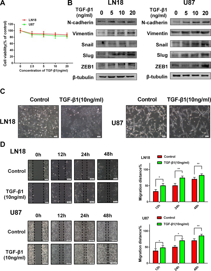
Figure 1. TGF-β1 induces an EMT-like process in GBM cells.
(A) MTT assay of cell viability in LN18 and U87 cells following exposure to TGF-β1 for 48 hours. (B) Western blot results of expressions of EMT-related proteins and transcription factors derived from LN18 and U87 cells treated with increasing concentration of TGF-β1 (0, 5, 10 and 20 ng/ml) for 48 hours. (C) The morphological changes of LN18 and U87 cells after exposure to TGF-β1 (10 ng/ml) for 48 hours under light microscope (×100 magnification). (D) Representative wound-healing images show migratory capacity in LN18 (×100 magnification) and U87 (×40 magnification) cells following exposure to TGF-β1 (10 ng/ml) compared with control group. Histograms show the mean level of migration distance observed in three random fields for each condition. *P < 0.05, **P < 0.01, control group versus TGF-β1 treated group for their respective time points.

