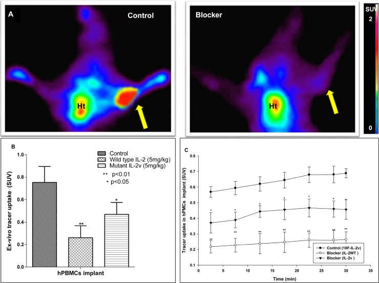
Figure 3. PET and ex-vivo biodistribution data of mutant [18F]FB-IL2v in SCID mice bearing an implant of PHA-activated human PBMCs (n = 4).
Control animals are compared to mice treated with 5 mg/kg (S.C) wild-type IL2 (IL2) or mutant IL2v prior to tracer injection. (A) Coronal PET images (25–30 min) of a control mouse and a mouse pretreated with wild-type IL2 (Blocker). The arrow indicates the location of the implant. Ht signifies the location of the heart. (B) Ex-vivo tracer uptake of mutant [18F]FB-IL2v in the PBMC implant 50 min after tracer injection. Tracer uptake was blocked with either wild-type IL2 or mutant IL2v. (C) PET-derived time activity curves (TACs) of mutant [18F]FB-IL2v in the implant of control mice and mice that were pretreated with wild-type or mutant IL2. Significant differences between the pretreated groups and the untreated controls are indicated by *(p < 0.05), **(p < 0.01).

