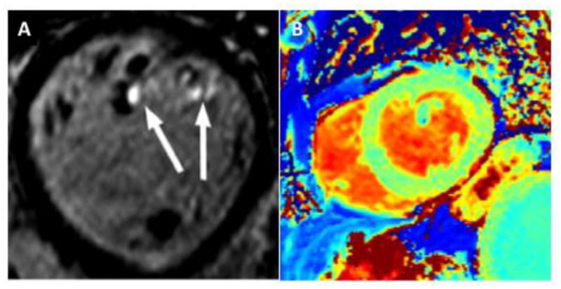
Figure 2.

Examples of focal and diffuse myocardial fibrosis in mitral valve prolapse (MVP). Short-axis view with three-dimensional late-gadolinium enhancement showing fibrosis of the papillary muscle tips (arrows) in a patient with bileaflet MVP and mild mitral regurgitation (A); precontrast T1 map with increased native T1 (1145 ms) indicating interstitial myocardial fibrosis in a different MVP patient with moderate-severe mitral regurgitation (B).
