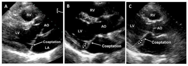
Figure 3.
Phenotypic spectrum of mitral valve prolapse (MVP). 2-dimensional echocardiographic parasternal long-axis images demonstrating (A) minimal systolic displacement, (B) anterior abnormal coaptation, and (C) posterior MVP. All show posterior leaflet bulging (arrows) relative to the anterior leaflet, but only MVP shows diagnostic superior leaflet displacement relative to the mitral annulus (dotted line) into the left atrium (LA). Posterior MVP and abnormal anterior coaptation are similar with regards to an increased coaptation height and an elongated posterior leaflet. Minimal systolic displacement shows posteriorly coapting leaflets, as seen in normal patients. AO = aorta; LV = left ventricle; RV = right ventricle.

