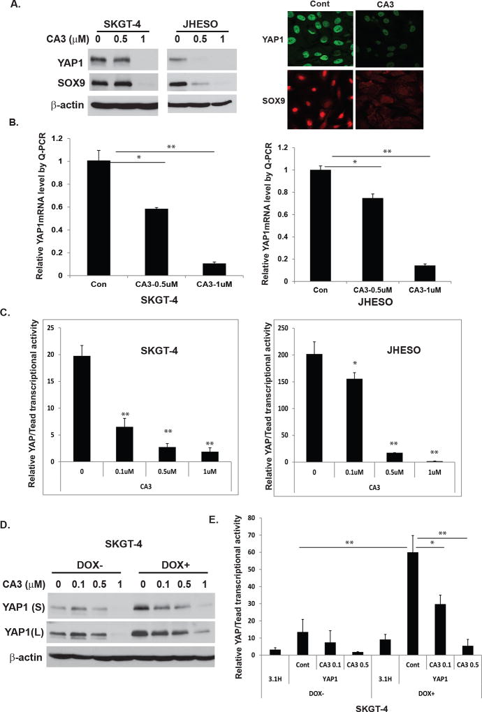
Figure 3. CA3 inhibits YAP1 expression and transcriptional activity in EAC cell lines, especially those with high YAP1.
A. Cell lysate from SKGT-4 and JHESO EAC cells treated with CA3 at indicated dosage were selected for immunoblotting analysis for YAP1 and SOX9 (left). Immunofluorescent staining of YAP1 and SOX9 in JHESO cells were observed by confocal microscopy (right). B. YAP1 mRNA levels were determined by quantitative real-time PCR in SKGT-4 and JHESO EAC cells treated with CA3 at indicated dosage. C. YAP1/Tead transcriptional activity was determined by co-transfection of Gal4-Tead and 5×UAS-luciferase and YAP1 cDNA in SKGT-4 and JHESO EAC cells and then treated with CA3 at indicated dosage. Luciferase reporter activity was measured after 48 h of transfection. For all experiments, values shown represent the mean and SD of at least triplicate assays (*P<0.05; **P<0.01). D&E. YAP1 expression (D) and transcriptional activity (E) were determined by immunoblotting or co-transfection of Gal4-Tead and 5×UAS-luciferase and YAP1 cDNA in SKGT-4 DOX− and DOX+ cells treated with CA3 at dosage indicated.

