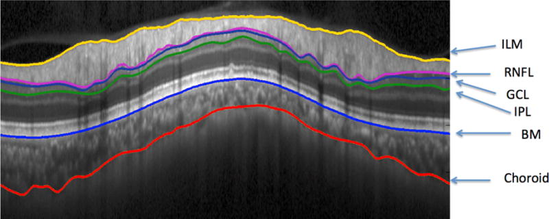
Figure 1.

Spectral-Domain Ocular Coherence Tomography (SD-OCT) image with San Diego Automated Segmentation Algorithm (SALSA) segmentation of the Internal Limiting Membrane (ILM), Retinal Nerve Fiber Layer (RNFL), Ganglion Cell Layer (GCL), Inner Plexiform Layer (IPL), Bruch’s Membrane (BM), and Choroidal-Scleral Interface
