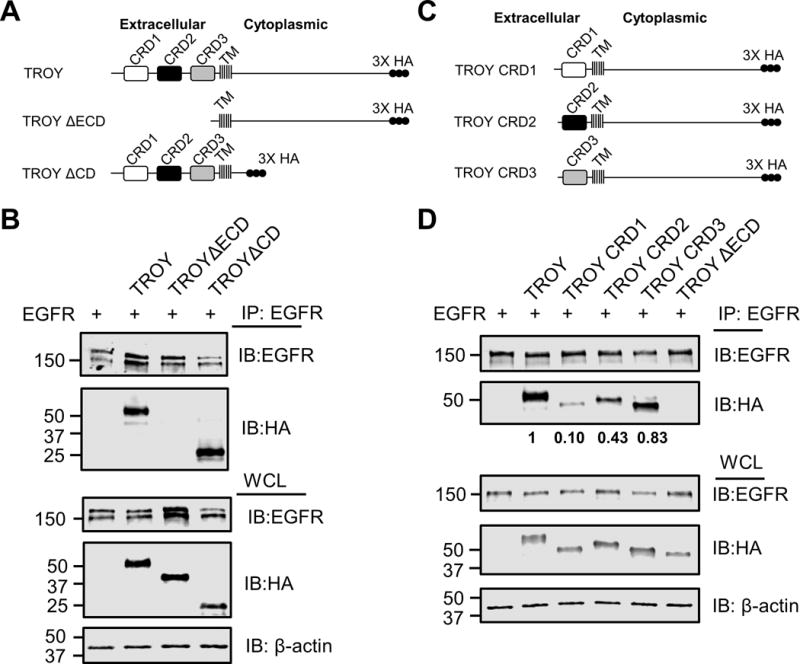
Figure 4.

TROY interacts with EGFR through its extracellular domain. (A) Schematic representation of the expression constructs encoding TROY WT, TROY ΔECD, or TROY ΔCD protein. The extracellular domain, transmembrane (TM) domain, and cytoplasmic domain are indicated and the 3X HA epitope tag is shown. (B) Q293 cells were co-transfected with EGFR and the indicated TROY constructs and 24 hr later cells were lysed and immunoprecipitated with anti-EGFR antibody. Immunoprecipitates or whole cell lysate (WCL) were immunoblotted (IB) with the indicated antibodies. The positions of molecular standards are shown on the left (in KDa). (C) Schematic representation of the expression constructs encoding TROY CRD1, or TROY CRD2 or TROY CRD3 protein. The transmembrane (TM) domain and cytoplasmic domain are indicated and the 3X HA epitope tag is shown. (D) Q293 cells were co-transfected with EGFR and the indicated TROY constructs and 24 hr later cells were lysed and immunoprecipitated with anti-EGFR antibody. Immunoprecipitates or whole cell lysate (WCL) were immunoblotted (IB) with the indicated antibodies. The positions of molecular standards are shown on the left (in KDa). The relative densitometry values of TROY variants in the anti-EGFR immunoprecipitates are shown.
