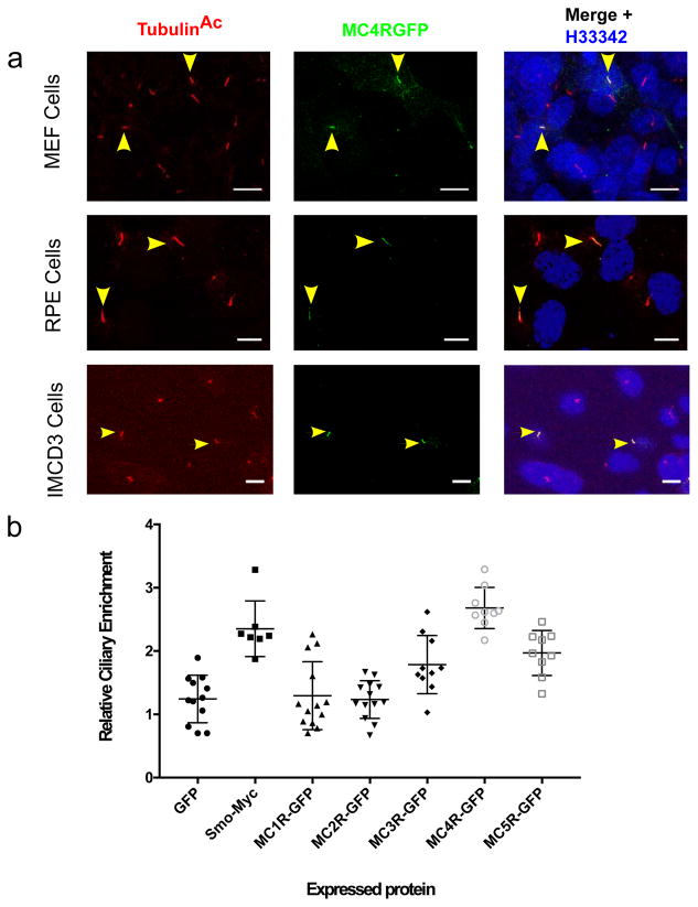
Figure 1. MC4R localizes to the primary cilia in heterologous cells.
A) Representative confocal microscopy images of transiently transfected MEF, RPE and IMCD3 cells, transfected with MC4R-EGFP labeled for the cilia specific protein acetylated Tubulin (TubulinAc, red) and GFP (green), and nuclei with Hoechst 33342 (blue). MC4R-GFP localize to the primary cilium (yellow arrowheads). Scale bars represent 10 μm. (B) Relative ciliary enrichment of melanocortin receptor family members compared to ciliary enrichment of GFP (negative control) and Smoothen (postive control). Data are Mean±sem. Means were compared to GFP (n=12 cells, mean=1.24) and Dunnet’s multiple comparison test was applied. Smo-Myc: n=7 cells, mean= 2.35, p=0,0001; MC1RGFP: n=13 cells, mean=1.29, p=0.9995, MC2RGFGP: n=13 cells, mean=1.23, p=0.9999, MC3RGFP: n=10 cells, mean=1.24, p=0.014; MC4RGFP: n=9 cells, mean=2.68, p=0.0001, MC5RGFP: n=9 cells, mean=1.96, p=0.0008.

