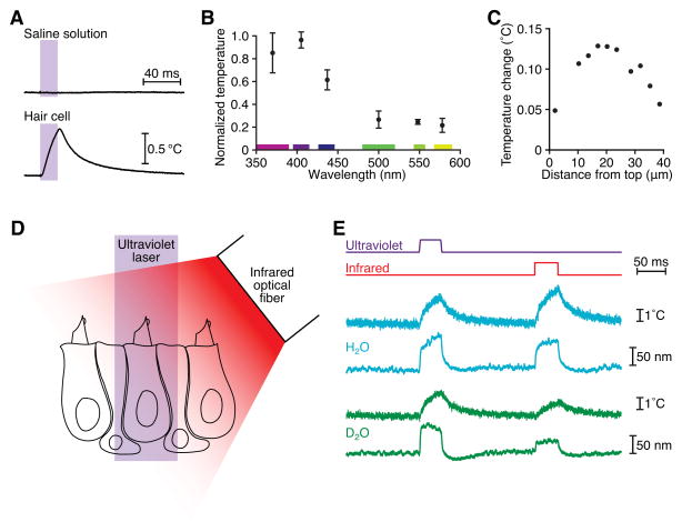
Figure 4. Thermal stimulation of hair cells.
(A) Ultraviolet irradiation of a hair cell led to heat generation, whereas irradiation of saline solution did not. This cell remained in the saccular epithelium and was irradiated from a direction orthogonal to the plane of the sacculus. The calibrated resistance of a microelectrode was used to record the temperature. (B) A temperature action spectrum reveals that heat production peaked in the 400 nm wavelength band. The means and standard deviations for four cells are plotted. The cells remained in the epithelium and were irradiated from a direction orthogonal to the plane of the sacculus. (C) Temperature measurements along a transect 2 μm from the lateral edge of a dissociated hair cell revealed that the largest temperature increases occurred basal to the hair bundle, whose top is situated at 0 μm and whose bottom is indicated by the dotted vertical line. This result accords with intra-mitochondrial light absorbers as the source of heat. (D) A schematic drawing depicts a hair bundle positioned beneath one beam of ultraviolet light and another of infrared light so that the bundle could be stimulated at either wavelength. (E) A hair bundle in H2O-based saline solution exhibited similar movements and local temperature increases (blue) for ultraviolet illumination at a power density of 71 MW·m−2 and for infrared irradiation at a power density of 0.13 MW·m−2. After substitution of D2O-based saline solution, the same hair bundle showed similar decreases in displacement and temperature change during infrared but not ultraviolet illumination (green). Temperature records were taken 2 μm from the side of the bundle, at a height of 7 μm above the apical epithelial surface.

