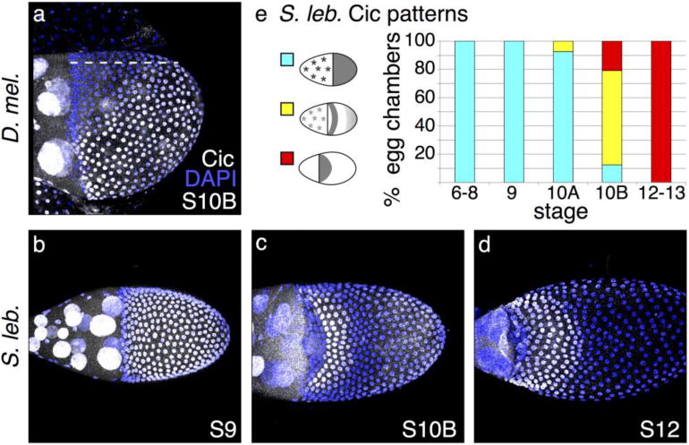
Fig. 4. Cic patterning confirms early EGF pathway discrepancies.

(a) Lateral view of D. melanogaster egg chamber; dotted line marks the dorsal midline. At S10B, Cic is in the nucleus of most follicle cells but clears from the nuclei of cells that lie above the dorsal anterior corner of the oocyte. (b) In early stages of S. lebanonensis oogenesis, Cic is present in all follicle cell nuclei, similar to early D. melanogaster Cic localization. (c) By S10B in S. lebanonensis, Cic is present in nuclei in a band of 4–5 rows of cells across the dorsal midline, 4 rows to the posterior of the anterior cortex. Nuclei of posterior follicle cells exhibit weak staining. (d) At S12, Cic remains present in the nuclei of anterior cells and has expanded to 7–8 rows. (e) Quantitation of S. lebanonensis Cic localization; N > 10 for each stage category.
