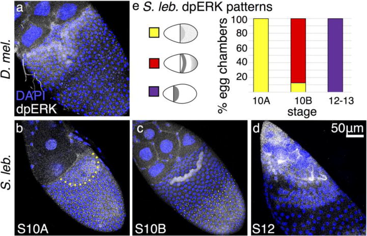
Fig. 5. dpERK patterning reveals early EGF-pathway discrepancies upstream of Cic.

(a–d) Anterior is to the upper left, dorsal is facing out of the page. (a) At S10B in D. melanogaster, dpERK is present most strongly in floor cells, with weaker localization in roof cells. (b) In S. lebanonensis, dpERK is present at S10A in a punctate pattern in most columnar follicle cells, but a rounded patch of dorsal anterior cells exhibit diffuse cytoplasmic staining; boundary marked by dashed line. (c) At S10B, the punctate dpERK pattern is restricted to the posterior follicle cells. Intense cytoplasmic dpERK staining appears in one band of follicle cells located 4– 5 rows to the posterior of the anterior cortex. (d) At S12, dpERK remains diffuse in anterior cells and has expanded to additional rows more posteriorly. (e) Quantitation of S. lebanonensis dpERK localization; N > 15 for each stage except for S12, where N=7.
