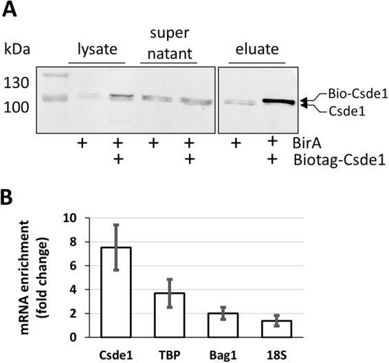
Figure 1.

Purification of Csde1 containing RNP complexes. (A) MEL cells expressing BirA, or expressing BirA plus biotag-Csde1 were lysed, and incubated with streptavidin beads. Supernatant and beads were collected, beads were washed and eluted. Western blot with fractions was stained with anti-Csde1 antibody. Size markers are indicated in kDa. The positions of endogenous and biotagged Csde1 are indicated. A single empty lane has been cropped. The uncropped image is available as Supplementary Figure S5. (B) RNA was isolated from eluates and tested for expression of Csde1, Tbp (Tata binding protein), Bag1 and 18S RNA by Q-PCR. The fold-change enrichment of the transcripts on streptavidin beads incubated with biotag-Csde1 lysate was calculated compared to pull downs from BirA MEL cells (error bars indicate SD, n = 3).
