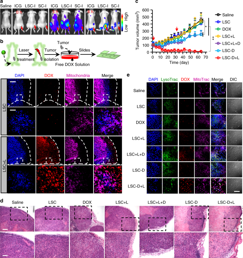
Fig. 7.
Overcoming cancer drug resistance and augmenting cancer destruction in vivo. a In vivo whole-animal imaging of indocyanine green (ICG) fluorescence at different times after intravenous injection via the tail vein in the form of free ICG, ICG-laden LSC (LSC-I) nanoparticles, and ICG-laden SC (SC-I) nanoparticles. The arrows indicate the locations of tumors in mice. b Fluorescence images of DOX in tumors of mice killed at 24 h post injection with LSC nanoparticles (with and without laser irradiation right before killing the mice). The data suggest the capability of overcoming cancer drug resistance could be achieved in vivo with the LSC + L treatment. The dashed lines indicate the boundary of the tumor tissue. The boxed regions are shown in higher magnification. Scale bar: 200 and 50 μm for low and high magnification images, respectively. c Growth curves of tumors in mice with various treatments showing augmented antitumor efficacy of the LSC-D + L treatments. Error bars represent s.d. (n = 7). **p < 0.01, *p < 0.05 (Kruskal–Wallis H-test). d Representative images of hematoxylin and eosin (H&E)-stained tumor tissue collected after killing the mice on day 64 after the first drug administration. Scale bar: 100 and 50 μm for low and high magnification images, respectively. e Fluorescence images of DOX in cells isolated form tumors collected on day 64 from mice with different treatments. The cells were incubated with DOX for 3 h. The data show that the drug-resistant capability of the multidrug-resistant cells in the tumors with the LSC-D + L treatment is still not fully recovered. Scale bar: 80 μm. All non-specified NIR laser (L) irradiation was at 1 W cm−2 for 2 min. Detached NCI/RES-ADR sphere cells enriched with CSCs were used to obtain xenograft tumors for all in vivo studies

